NOBLESVILLE, IN
Breast Thermography in Noblesville, IN
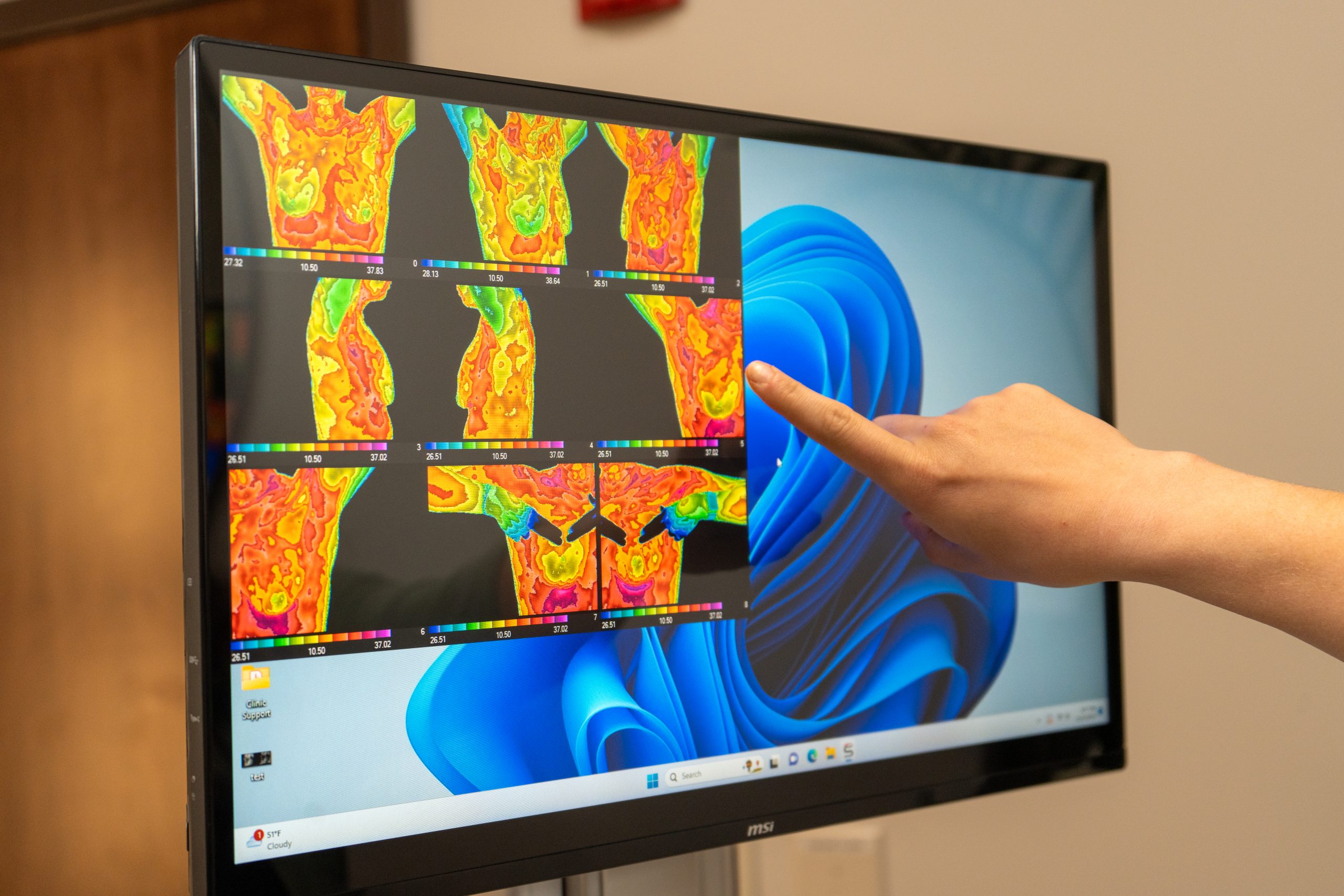
Breast thermography uses temperature-based imaging to identify early signs of abnormal tissue changes, offering a non-invasive way to monitor breast wellness.
NOBLESVILLE, IN
Breast thermography uses temperature-based imaging to identify early signs of abnormal tissue changes, offering a non-invasive way to monitor breast wellness.

Breast thermography is a non-invasive imaging technique used to help detect potential signs of breast cancer at an early stage.
This method relies on the fact that pre-cancerous and cancerous tissues typically exhibit elevated metabolic and vascular activity compared to healthy breast tissue. Because these abnormal cells require a steady flow of nutrients to support their growth, they often generate increased heat, which can be observed as higher surface temperatures during thermal imaging.

Thermography is a reliable screening tool with strong accuracy.

Best results come when thermography is used alongside mammography and clinical exams.

Thermography doesn’t show exact tumor location—it’s part of a bigger diagnostic picture.
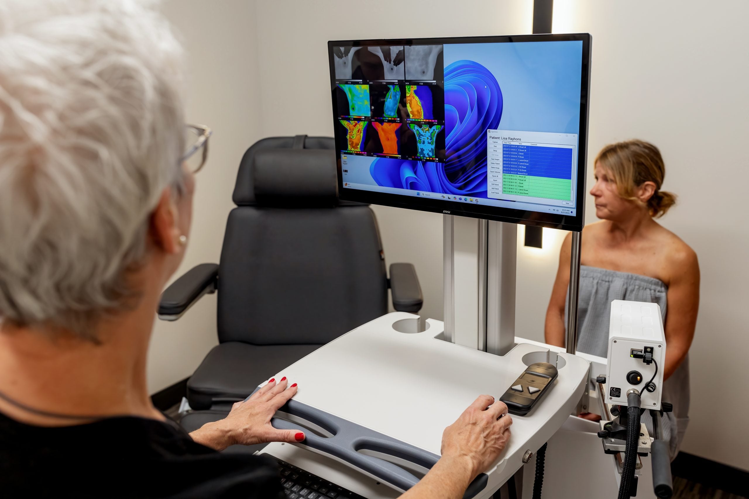
Thermography uses infrared (heat) sensors to detect heat and increased vascularity (angiogenesis) as the byproduct of biochemical reactions.
Completely safe: no radiation, non-invasive, and pain-free
Can be repeated as needed to monitor breast health, track treatment progress, or investigate concerns
Uses functional imaging to reveal physiological changes in breast tissue
No physical contact—imaging is done without touching the breast
Capable of detecting rapid changes in tissue earlier than traditional methods
Does not diagnose cancer but flags areas that may need additional evaluation
Can reveal disease-related changes in the breast up to a decade before tumors are typically found using other techniques
Particularly effective at identifying aggressive or fast-growing tumors through heat detection
A positive thermographic scan is one of the strongest indicators of potential future breast cancer—more significant than family history alone
Hormone therapies, including BHRT, do not affect the accuracy of results
High accuracy: approximately 90% sensitivity (detects 90% of cancers, with remaining cases often slow-growing or less invasive)
Around 90% specificity, reducing false positives and limiting unnecessary follow-up procedures
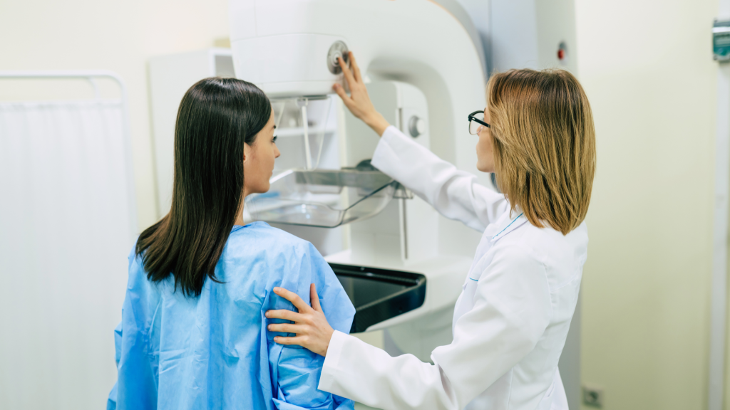
Mammography uses x-rays (radiation exposure) through the breast to produce a structural image. Areas of suspicion would need to be dense enough to be visualized.
Involves low-dose radiation for imaging purposes
Not recommended for frequent use due to radiation exposure and discomfort from breast compression
Identifies structural changes in breast tissue by analyzing density through X-ray imaging
Requires direct physical contact and compression of the breast for accurate results
Provides alerts for abnormalities but cannot confirm a cancer diagnosis on its own
Typically detects abnormalities after a tumor has already formed or symptoms have appeared
May not be effective in identifying fast-developing tumors in early, non-invasive stages
High rate of false positives: only about 9% of positive mammograms actually confirm disease presence
Hormone use—such as HRT—can reduce test sensitivity
Sensitivity averages around 80% in women over 50, but drops to about 60% for those under 60
Specificity averages 75%, leading to a higher chance of false positives—around 85% of biopsies following a mammogram return negative results
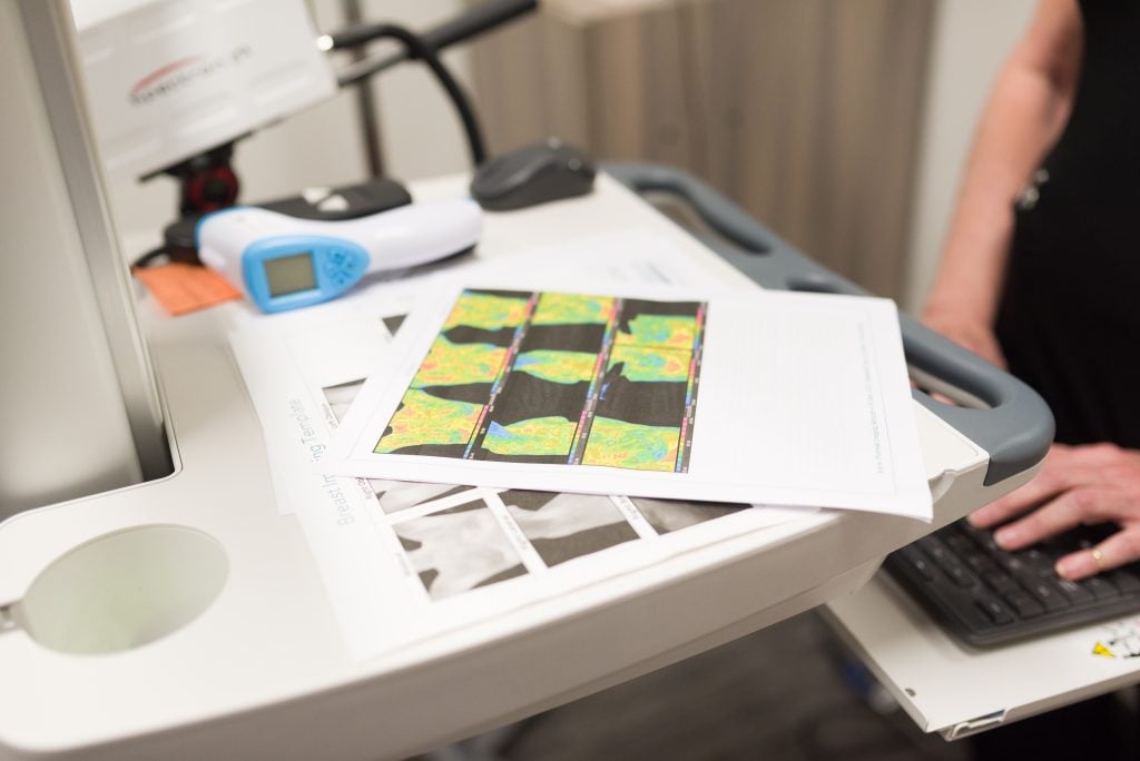
A trained thermography technician (RN or certified) will explain the procedure. An infrared camera and software are used to capture high-resolution images showing heat and vascular patterns in the breast tissue. Photos are taken from multiple angles, and only the thermographic images are saved and reviewed by a radiologist. The process is quick, painless, and involves no compression or radiation.



Well Infused Noblesville
Explore
Learn more
2025 © Well Infused LLC | All Rights Reserved | Well Infused trademarks and brands are the property of their respective owners