Get 10% Off Your First Service

Breast Thermography
Breast thermography is a diagnostic procedure that takes temperature-sensitive images of the breasts to aid in the early detection of concerning breast tissue changes.
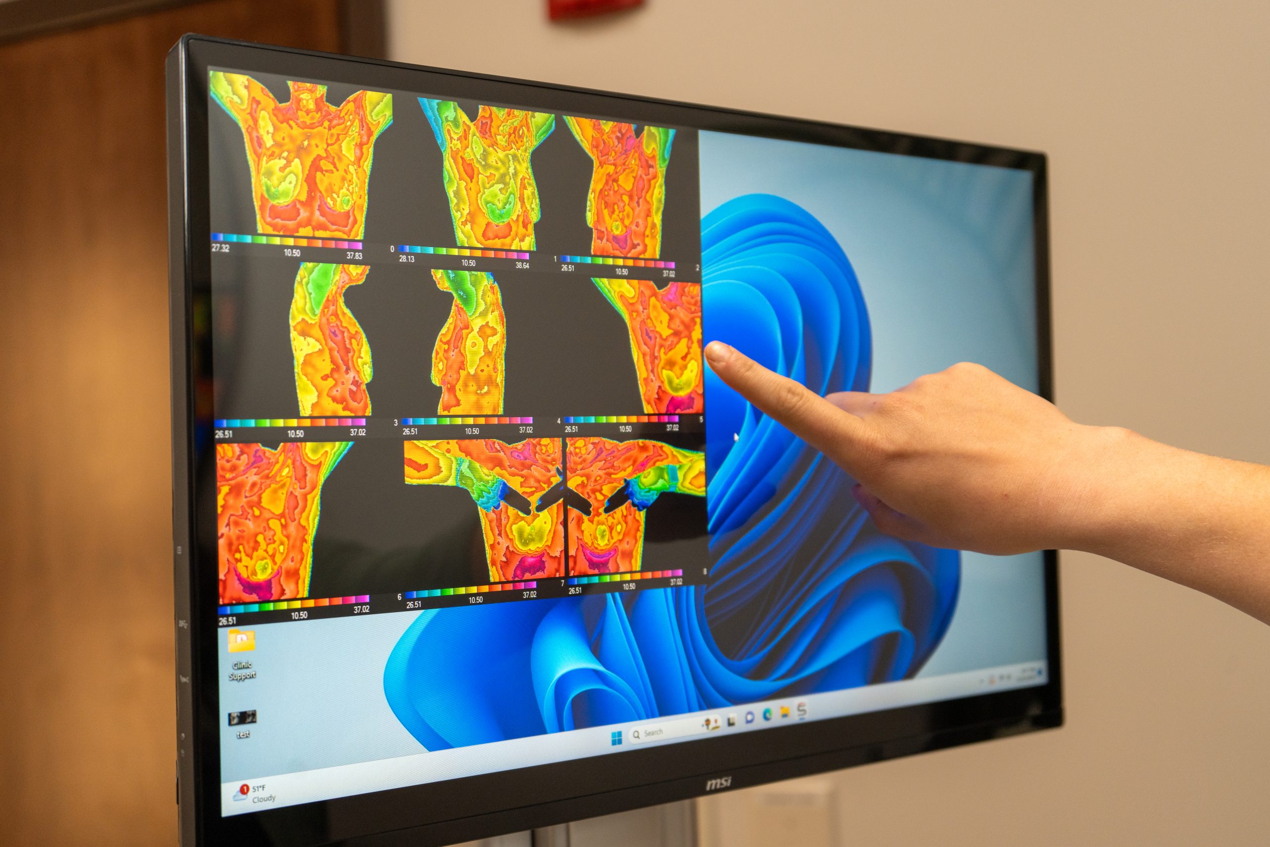
Breast thermography is a diagnostic procedure that takes temperature-sensitive images of the breasts to aid in the early detection of concerning breast tissue changes.

Breast thermography is a diagnostic procedure that images the breasts to aid in the early detection of breast cancer.
It is based on the principle that chemical and blood vessel activity in both pre-cancerous tissue and the area surrounding developing breast cancer is almost always higher than in the normal breast.
Since pre-cancerous and cancerous masses are highly metabolic tissues, they need an abundant supply of nutrients to maintain their growth, and this can increase the surface temperatures of the breast.

Thermography is a reliable screening tool with strong accuracy.

Best results come when thermography is used alongside mammography and clinical exams.

Thermography doesn’t show exact tumor location—it’s part of a bigger diagnostic picture.
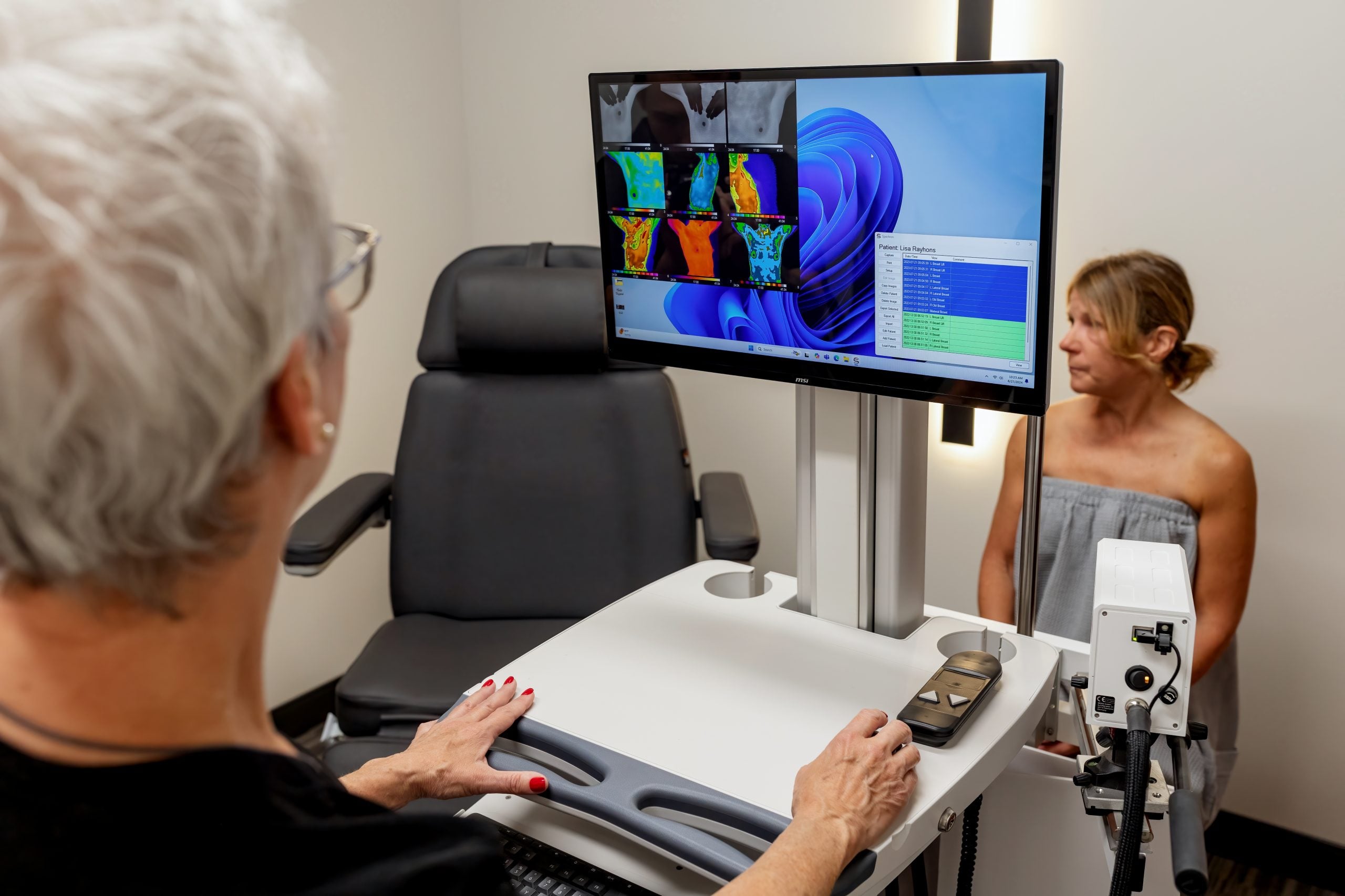
Thermography uses infrared (heat) sensors to detect heat and increased vascularity (angiogenesis) as the byproduct of biochemical reactions.
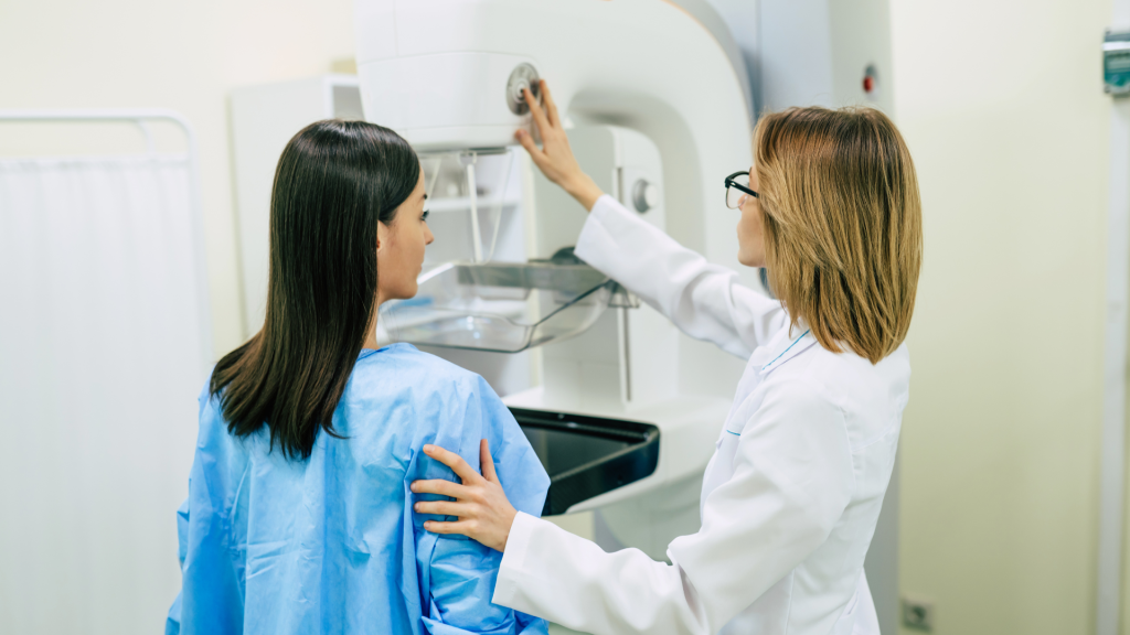
Mammography uses x-rays (radiation exposure) through the breast to produce a structural image. Areas of suspicion would need to be dense enough to be visualized.
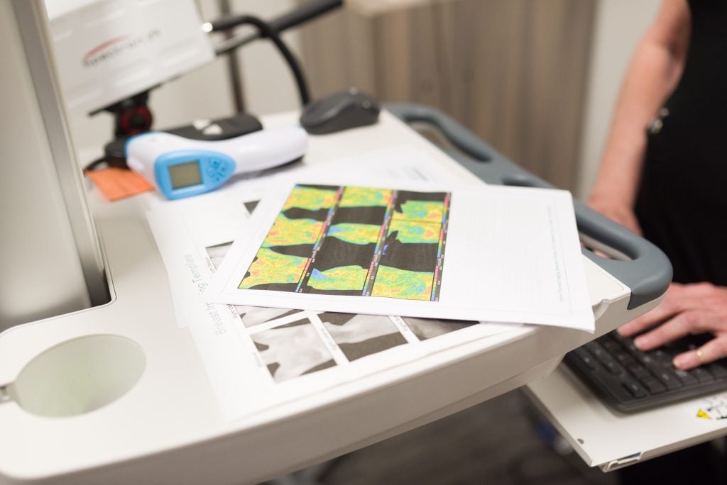
A trained thermography technician (RN or certified) will explain the procedure. An infrared camera and software are used to capture high-resolution images showing heat and vascular patterns in the breast tissue. Photos are taken from multiple angles, and only the thermographic images are saved and reviewed by a radiologist. The process is quick, painless, and involves no compression or radiation.